National Renal Pathology E.Q.A. Scheme
Circulation Z
This document gives information on individual cases in
circulation Z of this scheme. It contains no personal details
of participants.
Cases included:
Z 278
Z 279
Z 280
Z 281
Z 282
Z 283
End
A click on the  icon should provide an image from the material
circulated.
Some of the images are composites - remember to scroll the image
to see parts beyond the bottom of your screen.
icon should provide an image from the material
circulated.
Some of the images are composites - remember to scroll the image
to see parts beyond the bottom of your screen.
WARNING The image files associated with this
document are selected by the Organiser in an attempt to
illustrate the relevant features of the material which was
circulated in the EQA scheme. They are intended as an 'aide
memoire' for participants who may no longer have the slides for
review.
They are NOT intended as 'good examples' or as
teaching material. Some of the images may be chosen to
illustrate a feature which led some participants to a
wrong diagnosis.
Case Response Analysis
Circulation: Z
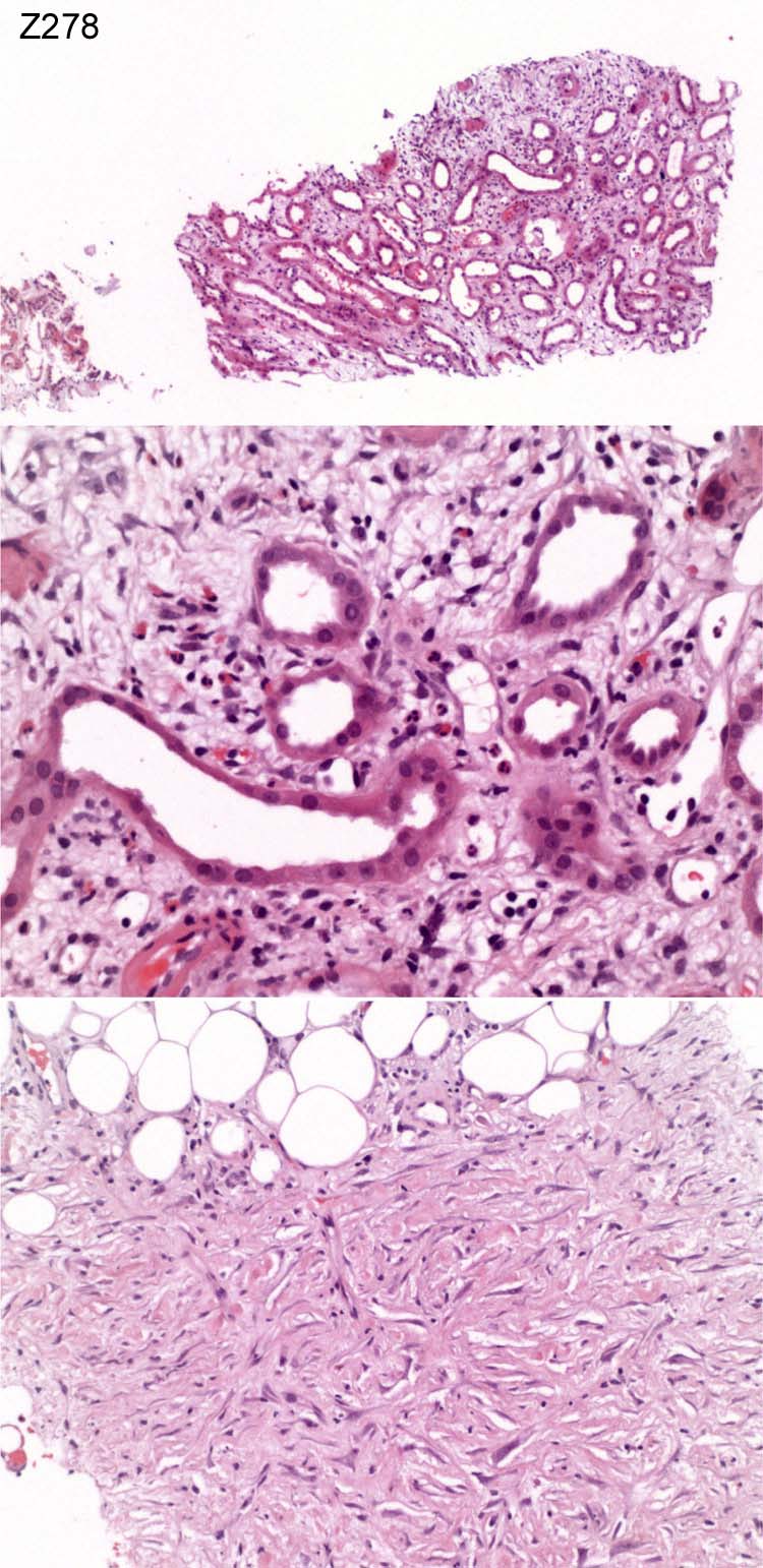
Case number: 278
....
Number of responses:82 . Date of analysis: 10 JAN 08
Clinical:
Male, 59 years old. E.coli UTI initially, given antibiotics. cANCA+ 1:80.
Creatinine 700 on admission. MPO PR3 negative. Now stuck at 400 ?cause.
Fibrin stain negative. Amyloid not present. Immunohistochemistry:
Negative for IgG, IgA, IgM and C3.
Specimen:
H&E
Diagnostic categories:                         Score:
 1  Specimen inadequate                                     1.83
 2  Kidney inadequate, odd fibrosis ?RPF in extrarenal fat? 1.34
 3  TIN & fat necrosis                                      0.49
 4  Tubulointerstitial nephritis                            2.56
 5  ATN                                                     1.10
 6  ATN with fibrosis in  extrarenal fat                    0.85
 7  Perinephric inflammation                                0.12
 8  Spindle celled tumour (+/- differential)                0.37
 9  TIN and inflammatory lesion in extrarenal fat           1.34

Asterisks (if any) indicate dangerous diagnoses.
Highest scoring diagnosis was 4 with 2.56
Secondary diagnoses and comments (if any):
Unsuitable for EQA*42. Needs imaging to exclude tumour*3. Drug induced?*3.
Exclude ascending infection*2. Immuno. to sort out spindle cell
proliferation*1. Wrong slide sent?*1.
Original report and further information (if any):
Renal biopsy inadequate, possible interstitial nephritis, but peri-renal
tissue shows fibrosis suggesting possible retroperitonela fibrosis.
Subsequent enquiries revealed that the patient had had an aorto-femoral
bypass. No other explanation for the fibrosis is available. Nothing to
suggest ureteric obstruction
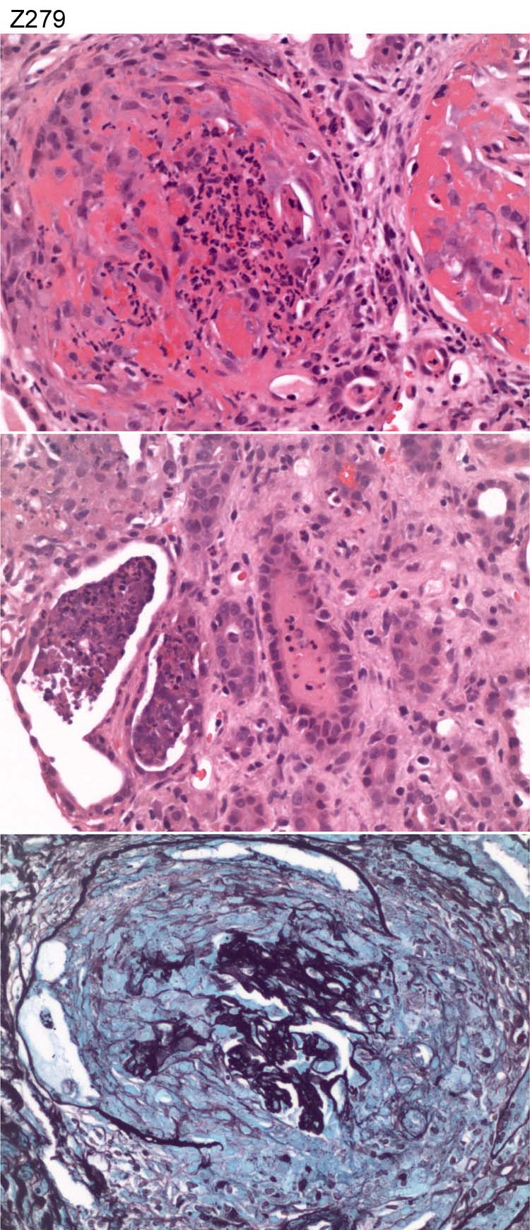
Circulation: Z
Case number: 279
....
Number of responses:82 . Date of analysis: 10 JAN 08
Clinical:
Female, 45 years. History of loose motions for one week. Hypertensive.
Acute deterioration in renal function with urea >40 and creatinine of 1600.
Anuric at presentation. Urine dipstick protein +++, blood +++. Anti GBM Ab
positive by quick card test. No fluorescence sample sent.
Specimen:
H&E & Meth Silver
Diagnostic categories:                         Score:
 1  Crescentic Gn c/w Goodpasture's                         9.88
 2  Crescentic Gn NOS                                       0.12

Asterisks (if any) indicate dangerous diagnoses.
Highest scoring diagnosis was 1 with 9.88
Secondary diagnoses and comments (if any):
ANCA*10. Exclude UTI*2. EM*3. MSB*1. ATN too*1. Immnoperoxidase*2.
Hypertensive vascular disease*5. Arteriolitis-?part ot anti-GBM??
hypertensive ?ANCA*7. Are those megakaryocytes?*1. Exclude acute
pyelonephritis*2.
Original report and further information (if any):
Necrotising glomerulonephritis with 100% crescents, consistent with anti-
GBM disease.
Circulation: Z
Case number: 280
....
Number of responses:82 . Date of analysis: 10 JAN 08
Clinical:
Male, 77 years old. Acute renal failure (Creatinine 1770). History of
bladder carcinoma, hypertension and hypercholesterolaemia. Raised CRP (
100). Light chains present in urine. CT scan showed peculiar streaking of
perinephric fatty tissue highly suspicious for acute pyelonephritis
radiologically. Immunofluorescence negative for all stains. Amyloid stains
negative. EM non-contributory.
Specimen:
H&E
Diagnostic categories:                         Score:
 1  Tubulointerstitial nephritis                            0.49
 2  Tubulointerstitial nephritis AND atheroembolus          0.98
 3  Myeloma cast nephropathy and cholesterol emboli         3.29
 4  Atheroemboli                                            0.73
 5  Myeloma cast nephropathy (+/- TIN)                      2.05
 6  Acute TIN AND atheroemboli AND myeloma                  2.07
 7  Atheroemboli and pyelonephritis                         0.12
 8  Light chain deposition disease                          0.12
 9  Myeloma, acute pyelonephritis & cholesterol emboli      0.12
 10 Pyelonephritis                                          0.02

Asterisks (if any) indicate dangerous diagnoses.
Highest scoring diagnosis was 3 with 3.29
Secondary diagnoses and comments (if any):
Drug history*4. Exclude myeloma*8. K & L*5. Exclude infection*10.
Hypertensive vascular disease*4. Levels*1. EVG*1. Exclude neoplastic
lymphoid infiltrate*6. Too complex/unsuitable for EQA*3.
Original report and further information (if any):
Acute-on-chronic tubulo-interstitial nephritis, with possible myeloma
casts. Rule out ascending infection.
Subsequently a bone marrow revealed an increase in plasma cells and a
diagnosis of myeloma was made. The patient died.
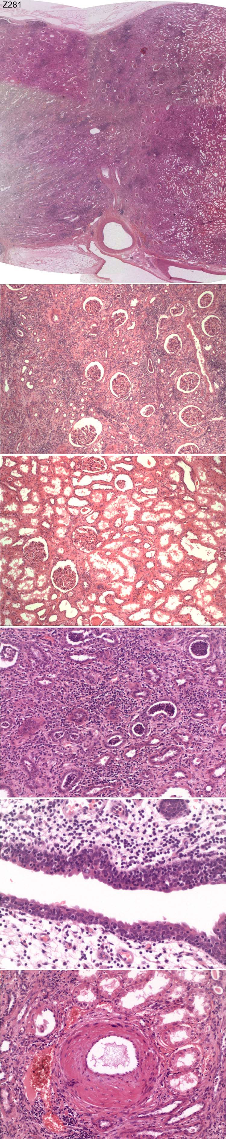
Circulation: Z
Case number: 281
....
Number of responses:82 . Date of analysis: 10 JAN 08
Clinical:
Male, 42 years old. Progressive deterioration in renal function to end
stage. Poorly controlled hypertension. Nephrectomy. Subsequent pan-
hypopituitarism (ZN negative. PAS unremarkable).
Specimen:
H&E
Diagnostic categories:                         Score:
 1  Reflux / chronic pyelo. + acute infection               2.32
 2  Reflux nephropathy / chronic pyelonephritis             1.59
 3  Granulomatous TIN / Sarcoid                             2.80
 4  Tubulointerstitial nephritis                            0.55
 5  Acute pyelonephritis                                    0.30
 6  Segmental hypoplasia                                    0.12
 7  Acute pyelonephritis and sarcoidosis                    0.12
 8  Granulomatous pyelonephritis                            1.95
 9  Hypertensive / ischaemic damage                         0.18
 10 Lymphoma                                                0.06

Asterisks (if any) indicate dangerous diagnoses.
Highest scoring diagnosis was 3 with 2.80
Secondary diagnoses and comments (if any):
Exclude TB*5. Investigations for sarcoid*13. ?drugs*2. ?HbS*2. Papillary
necrosis*1. Need to see whole kidney*3.
Original report and further information (if any):
1) Acute on chronic pyelonephritis (? reflux-related)
2) Granulomas, raising the possibility of sarcoidosis. ZN negative.
Circulation: Z
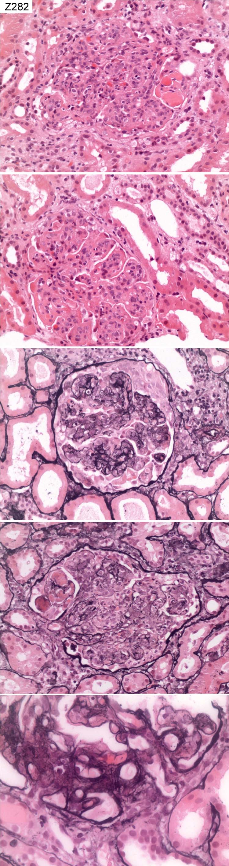
Case number: 282
....
Number of responses:82 . Date of analysis: 10 JAN 08
Clinical:
Female, 39 years old. ?Lupus nephritis. Special stain for amyloid
negative. Immunohistochemistry: Mesangial and membrane deposition for IgG,
IgA, IgM and complement component C3 and C9.
Specimen:
H&E, PAS
Diagnostic categories:                         Score:
 1  Lupus class IV G - A                                    6.83
 2  Lupus class IV NOS                                      2.20
 3  Lupus class IV A/C                                      0.49
 4  Lupus class Vd                                          0.12
 5  Necrotising Gn?lupus                                    0.12
 6  Membranoproliferative, c/w Class IV lupus               0.24

Asterisks (if any) indicate dangerous diagnoses.
Highest scoring diagnosis was 1 with 6.83
Secondary diagnoses and comments (if any):
Lupus serology?*6. C1q immuno?*5. EM*10. Exclude cryo*2. Anti-C3Nef.*1.
Original report and further information (if any):
Lupus class IV, active, no sclerosis, diffuse.
Circulation: Z
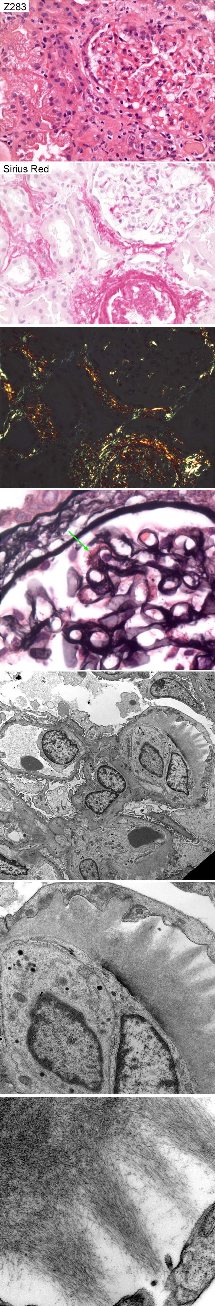
Case number: 283
....
Number of responses:82 . Date of analysis: 10 JAN 08
Clinical:
Female, 82 years old. Nephrotic syndrome ?cause. Normal renal function.
IgG lambda paraprotein4 g/l. IMF negative.
Specimen:
H&E, Silver and Sirius Red. Photo's of EM x 3
Diagnostic categories:                         Score:
 1  Amyloidosis                                             10.00

Asterisks (if any) indicate dangerous diagnoses.
Highest scoring diagnosis was 1 with 10.00
Secondary diagnoses and comments (if any):
Presumably AL type *21. Investigate amyloid type*8. Exclude simultaneous
pyelonephritis*1. Exclude myeloma*8.
Original report and further information (if any):
Amyloidosis. Subsequent demonstration of monoclonal (lambda) plasma cell
population in bone marrow.
Links to cases in this document:
Top
Z 278
Z 279
Z 280
Z 281
Z 282
Z 283
Last updated: 10 JAN 08
Organiser:
Professor Peter Furness, PhD, FRCPath.
Department of Pathology
Leicester General Hospital
Gwendolen Road
Leicester
LE5 4PW, U.K.
Tel: (0116)2584582
Fax: (0116) 2584582
Email:
peter.furness@le.ac.uk